Thursday 29 September 2022

How to examine an ulcer? - A brief review.

 An ulcer is break in the continuity of covering epithelium- skin or mucous membrane. It may either follow molecular death of the surface epithelium or its traumatic removal.


History:

In any medical cases, if you want to find the solid cause of disease, you need a clear history about particular symptoms or disease.

The following points noted in history of case of an ulcer:
  • Mode of onset
  • Duration
  • Pain
  • Discharge
  • Associated disease.

Mode of onset- 

How and when the ulcer developed? After some injury or spontaneously?
1.If ulcer occurs because of trauma- on removal of traumatic agents it will get healed.
e.g. Traumatic injuries of oral mucosa due to sharp food stuffs.
2.Ulcer which originate spontaneously may follow swelling, which may be matted tuberculous lymph nodes or gumma or rapidly growing malignant tumour. e.g. epithelioma or malignant melanoma.

Duration-

How long ulcer present there?
Acute ulcer- short duration
Chronic ulcer- long duration.
Incubation period should also known i.e time interval between exposure & onset of ulcer. example: in syphilis- 3 to 4 weeks, chancroid (soft sore) - 3 to 4 days.

Pain-

Ulcer which associates with inflammation will be painful.
Syphilitic ulcer & tropic ulcer resulting from nerve diseases (tabes dorsalis, transverse myelitis, peripheral neuritis) are painless.
Tuberculous ulcers are slightly painful.
Ulcers from malignant diseases such as epithelioma or basal-cell carcinoma are initially painless, becomes painful if infiltrate structure supplied by pain nerve endings.

Discharge-

If it discharges enquiry must be made about its nature- serum, pus or blood.

Associated disease-

Nervous diseases like tabes dorsalis, syringomyelia, transverse myelitis & peripheral neuritis result an ulcer (trophic or perforating ulcer). 
Generalized tuberculosis, nephritis or diabetes may lead to ulcer formation.
Syphilis at primary stage gives rise to chancre and in tertiary stage rise to a gummatous ulcer.

Physical examination:

In case of ulcer, physical examination plays major role, because ulcer may be a sequel of malnutrition, general atherosclerosis, syphilis, tuberculosis etc.

General examination-

1. If ulcer is suspected to be syphilitic, a thorough search should be made for presence of other syphilitic stigmas in the body.
2. If it appears to be tuberculous, all lymph nodes in the body should be examined along with other examination such as chest, the neck, the abdomen etc.
3. If ulcer seems to be due to atherosclerotic or buerger's disease (ischaemic), the whole body must be examined for presence of atherosclerosis or its complication anywhere in the body. Moreover buerger's disease is bilateral condition and the other limb should always be examined.
4. When the ulcer is a trophic (perforating) one general examination must be made to know the type of nervous disease present with this condition.

Local examination:

It includes Inspection, Palpation, Examination of lymph nodes, Examination for vascular insufficiency, Examination for nerve lesion.

A. INSPECTION-

1. Size and Shape

Tuberculous ulcer

Oval in shape, irregular crescentic border.

Syphilitic ulcers

Circular or semilunar to serpiginous ulcer

Varicose ulcers

Vertically oval in shape

Carcinomatous ulcers

Irregular in shape & size


A bigger ulcer will definitely take longer time to heal.
How to record exactly the size & shape of the ulcer- A sterile gauge may be pressed on to ulcer to get its measurements.
2. Number-
Tuberculous, gummatous, varicose ulcers & soft chancres may be more than one in number.

3. Position-
Varicose ulcer- medial malleolus of lower limb which shows varicose veins.


Rodent ulcer- confined to upper part of the face above line joining the angle of the mouth to the lobule of ear, occuring frequently in inner canthus of eye.


Tuberculous ulcers- neck & axilla or groin.
Lupus- form of cutaneous tuberculosis occurs more frequently on face, fingers & hand.
Hunterian chancre & soft sores- found over the external genitalia.
Gummatous ulcers- subcutaneous bones such as tibia, sternum, skull etc.
Perforating or trophic ulcers- heel of foot or ball of the foot, which carries maximum weight of the body.
Malignant ulcers- occur anywhere in body but most commonly seen on lips, tongue, breast, penis & anus.


4.Edge- 
Edge should not be confused with margin.
Edge is area between margin & floor of ulcer. Margin is junction between normal epithelium & ulcer.

Spreading ulcer

Edge is inflamed & oedematous

Healing ulcer

Shows blue zone (due to thin growing epithelium)

White zone (due to fibrosis of the scar)


Five common types of ulcer edge:


Undermined edge

Tuberculosis (ulcer spreads in & destroys the subcutaneous tissue faster than it destroys the skin, overhanging skin is thin, friable, reddish blue and unhealthy.

Punched out edge

Gummatous ulcer or deep trophic ulcer. (ulcers are limited to ulcer itself & do not tend to spread to surrounding tissue.)

Sloping edge

Healing traumatic or venous ulcers.

Raised & pearly white beaded edge

Rodent ulcer (this type develops in invasive cellular disease & becomes necrotic at the centre)

Rolled out (everted) edge

Squamous celled carcinoma or ulcerated adenocarcinoma.

 5. Floor-

Exposed surface of ulcer.
If floor is covered with red granulation tissue, ulcer seems to be healthy and healing.
Pale and smooth granulation tissue indicates slow healing ulcer. 
Wash-leather slough (like wet chamois leather) on the floor of an ulcer is pathognomonic of gummatous ulcer.
Black mass on floor suggests malignant melanoma.
Trophic ulcer penetrates down to bone which forms floor.

6. Discharge-
Amount and smell of the discharge should be noted.
Healing ulcer- scanty serous discharge.
Spreading & inflamed ulcer- purulent discharge.
Tuberculous or malignant ulcer- sero-sanguineous discharge.
B-pyocynea infection- greenish discharge.
[Always advisable to take bacteriological wab of the ulcer]

7. Surrounding area-
In acute inflammation- surrounding area of an ulcer is glossy, red & oedematous.
In varicose ulcer- eczematous & pigmented.
Old case of tuberculosis- scar or wrinkling in surrounding skin of an ulcer.

8. Whole limb should be examined in case of ulcer. 
Presence of varicose vein and deep vein thrombosis will indicate the ulcer to be varicose ulcer.
Neurological insufficiency will indicate the ulcer to be trophic one.

B. PALPATION-

1. Tenderness:
Acutely inflamed ulcer - more tender.
Chronic ulcers like tuberculosis and syphilitic ulcers - slightly tender.
2. Edge & margin:
Activity is maximum at the margin & edge of the ulcer.
Marked induration (hardness) of edge is feature of carcinoma.
A certain degree of induration or thickness is expected in any chronic ulcer.
3. Base:
You should know the difference between base & floor.
Base is on which ulcer rests, Floor means exposed surface within the ulcer.
Marked induration of the base is important feature of squamous celled carcinoma & hunterian chancre.
4. Depth:
It can be recorded in millimeter, Trophic ulcers may be as deep as to reach the bone.
5. Bleeding:
Bleeding on touch is common feature of malignant ulcer.
6. Relation with deeper structure:
A gummatous ulcer- over subcutaneous bone often fixed to it.
Malignant ulcer fixed to deeper structure by infiltration.
7. Surrounding skin:
Increased tenderness & temperature of surrounding skin indicates ulcer to be of acute inflammatory origin.
Fixity to deeper structure- malignant nature of lesion, surrounding skin is tested for nerve lesion (loss of sensation or motor deficit).

C. Examination of lymph nodes: [most important part of examination]

In acutely inflamed ulcers- regional lymph node enlarged, tender, & show signs of acute lymphadenitis.
Tuberculous ulcer- lymph nodes are enlarged matted & slightly tender.
Hunterian chancre- lymph node remains discrete, firm & shotty. (pathognomonic of hard [hunterian] chancre).
Gummatous ulcers- lymph node usually not invloved.
In rodent ulcer- lymph node not invloved because early obliteration of lymphatics by neoplastic cells.
In malignant ulcer- nodes are stony hard and may be fixed to neighbouring structures in late stage.
Stony hard consistency- suggest secondary invlovement.

D. Examination of vascular insufficiency:

The clinician must examine the condition of arterial proximal to the ulcer.
Atherosclerosis, buerger's disease, Raynaud's disease etc. may be cause of ulcer from poor circulation.

E. Examination for nerve lesion:

Trophic ulcers - develops due to repeated trauma to insensitive part of patient's body. Mostly seen in sole, as this is weight bearing zone if there is sensory loss.

Special investigation:

1. Routine examination of blood.
2. Examination of the urine.
3. Bacteriological examination of discharge.
4. Skin test.
5. Chest X ray.
6. Biopsy.
7. X-ray of the bone & joint.
8. Contrast radiography.
9. Imaging technique.

Saturday 24 September 2022

Development , histology and clinical anatomy of tongue

Development of tongue: 

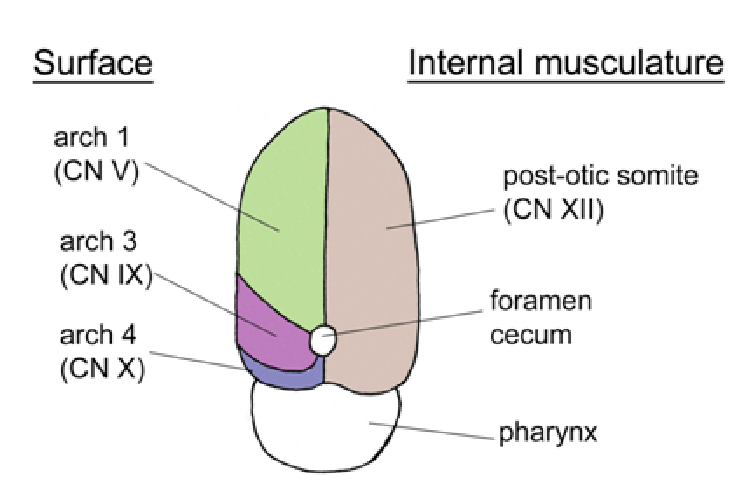
The tongue develops in relation to ventral portion of the pharyngeal arches in the floor of developing mouth. Primordia of tongue seen in embryo of 4 weeks.

Primordia of developing tongue:

Lateral lingual swellings- Medial most parts of first pharyngeal (mandibular) arches proliferate to form two lateral swellings.
Tuberculum impar - Also formed by proliferation of median most part of 1st pharyngeal arches. It is median swelling which partially separates two lateral lingual swelling.


Hypobranchial eminence- another median swelling formed by mesoderm of 2nd, 3rd, and part of 4th pharyngeal arches. It divides into two parts:
       Cranial part (copula) which is related to 2nd & 3rd arches.
       Caudal part, which is related to 4th arch. It forms the epiglottis. Immediately behind this swelling is the laryngeal orifice, which is flanked by the arytenoid swellings.

Formation of tongue:


Anterior two-thirds of tongue [derived from first pharyngeal arch] -  
                                               ➤  Two lateral lingual swelling increase in size, merge with each other, overgrow the tuberculum impar.
                                               ➤ The merged lateral lingual buds form the anterior two-thirds of the tongue.
                                               ➤ Fusion of the two lateral lingual buds is indicated by middle groove, the median sulcus of tongue.

Posterior one-thirds of tongue [formed by copula-cranial part of hypobranchial eminence] -
                                                ➤The 3rd arch mesoderm of copula grows over the 2nd arch mesoderm of copula and fuses with mesoderm of 1st arch.
                                                ➤ The V shaped groove separates the anterior two-thirds of the tongue from the posterior one third.


Most posterior part of tongue and epiglottis -
                                                ➤   Formed by caudal part of hypobrachial eminence derived from 4th arch mesoderm 
  
Musculature of the tongue -
                                                ➤ Mainly derived from myoblasts originating in occipital somites. Tongue musculature is innervated by hypoglossal nerve.  
     

Histology of tongue: 

  1. The bulk of tongue made up of striated muscles.
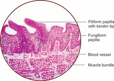
  2. The mucous membrane consists of a layer of connective tissue (corium), lined by stratified sqamous epithelium. 
  3. On the oral part of the dorsum, it is thin, forms papillae & adherent to the muscles.
  4. On the pharyngeal part of the dorsum, it is rich in lymphoid follicles.
  5. On the inferior surface, it is thin and smooth. Numerous glands, both mucous & serous lie deep to the mucous membrane.

Taste buds: numerously present on side of vallate papillae, on wall of surrounding sulci, foliate papillae, and over the posterior one-third of tongue, sparsely on fungiform papillae, soft palate, epiglottis and pharynx.
No taste buds on mid-dorsal region of oral part of tongue.


Thursday 22 September 2022

Muscles of tongue( why you can't swallow the tongue itself?😂), arterial supply, venous and lymphatic drainage, nerve supply.

Tongue

It is a muscular organ, situated in floor of mouth, comprises voluntary skeletal muscle. It appears very mobile, still it cannot be swallowed like food, we will discuss about it later. It has 4 extrinsic & intrinsic muscle. Out of which 1 extrinsic muscle (palatoglossus) supplied by vagoaccesory complex. Remaining muscle supplied by hypoglossal nerve. In paralysis of hypoglossal nerve, on protrusion tip of the tongue deviates to paralysed side.

External features:

It has 
a. Root
b. Tip
c. Body (curved upper surface[dorsum] & inferior surface)

a) Root:


b) Tip of the tongue- forms the anterior free end which at rest, lies behind the upper incisor teeth.
c) Curved upper surface(dorsum)- divided into oral & pharyngeal parts.
    Inferior surface- confined to oral part only.

Dorsum of tongue:


Dorsum of tongue is convex in all direction, divides into two part oral & pharyngeal.
An oral part or anterior two thirds: 
  • Also called papillary part of the tongue, placed on floor of the mouth. Margin contacts with the gums and teeth.
  • In front of the palatoglossal arch, each margin shows a 4 to 5 vertical folds, called foliate papillae
  • Superior surface of oral part covered with papillae which make it rough.
  • Inferior surface is smooth and it shows a median fold called frenulum linguae.
  • On either side of frenulum there is prominence produced by deep lingual vein.

The pharyngeal or lymphoid part of the tongue or posterior one-third
  • Divides from anterior part of tongue by V shaped groove called sulcus terminalis. The meeting point of V called foramen caecum.
  • The most posterior part called base of tongue, forms the anterior wall of the oropharynx.
  • Mucous membrane has many lymphoid follicles, that collectively constitute lingual tonsil, it also contain mucous gland.

Papillae of the tongue:


Vallate or circumvallate papillae

Fungiform papillae

Filiform papillae or conical papillae

Foliate papillae

o   Larger in size and 8-12 in number.

o   Situated in front of sulcus terminalis.

o   Numerous near the tip & margin of tongue.

o   Some are scattered over the dorsum.

o   Smaller than vallate papillae but larger than the filiform papillae.

o   Bright red in color.

o   Covers presulcal area of the dorsum of tongue.

o   It has characteristic velvety appearance.

o   They are smallest and most numerous.

o   Few foliate are present.


Muscles of the tongue:

Middle fibrous septum divides the tongue into right and left halves. Each half contains 4 intrinsic and extrinsic muscles.


Intrinsic muscles

Extrinsic muscles

Superior longitudinal

Inferior longitudinal

Transverse

Vertical

Genioglossus

Hyoglossus

Styloglossus

Palatoglossus.


Intrinsic muscleoccupies the upper part of the tongue. Attached to submucous fibrous layer and to median fibrous septum. They alter the shape of tongue.


Intrinsic muscles

Location

Function

Superior longitudinal

Lies beneath the mucous membrane

Shortens the tongue makes the dorsum concave.

Inferior longitudinal

Lying close to inferior surface of tongue between the genioglossus & hyoglossus

Shortens the tongue makes the dorsum convex.

Transverse muscle

Extend from median septum to margin.

Makes tongue narrow and elongated.

Vertical muscle

Found at the border of anterior part of tongue

Makes tongue broad and flattened.


Extrinsic muscles: connect the tongue to the mandible via genioglossus: to the hyoid bone through hyoglossus: to the styloid process via styloglossus, and palate via palatoglossus.

Extrinsic muscle

Origin

Insertion

Action

Palatoglossus

Oral surface of palatine aponeurosis

Descends in palatoglossal arch to side of tongue

Pulls up the root of tongue, approximates the palatoglossal arches and thus closes the oropharyngeal isthmus.

Hyoglossus

Whole length of greater cornua and lateral part of hyoid bone

Side of tongue between styloglossus and inferior longitudinal muscle of tongue.

Depresses tongue,

Makes dorsum convex,

Retracts protruded tongue.

Styloglossus

Tip and part of anterior surface of styloid process

Into side of tongue

Pulls tongue upwards and backwards.

Genioglossus

Upper genial tubercle of mandible

Upper fibres into tip of tongue, middle fibres into dorsum, lower fibres into hyoid bone.

Retracts the tongue,

Depresses the tongue,

Pulls posterior part of tongue forwards and protrude the tongue forwards. Life- saving muscle.


Arterial supply of tongue:

Derived from tortuous lingual artery a branch of external carotid artery.
The root of the tongue supplied by tonsillar, a branch of facial artery, and ascending pharyngeal, branch of external carotid.

Venous drainage:

Two venae comitantes accompany the lingual artery and one vena comitantes accompanies the hypoglossal nerve.
The deep lingual vein -
  • The largest and principle vein of the tongue.
  • Course: visible on inferior surface of the tongue, runs backwards and crosses the genioglossus and hyoglossus below the hypoglossal nerve.

Lymphatic drainage:


Tip of the tongue

Drains bilaterally to the submental nodes.

Right & left half of remaining anterior 2/3rd

Unilaterally to submandibular nodes.

Posterior one-third

Bilaterally to the jugulo-omohyoid nodes[lymph node of tongue].

Posterior most part

Bilaterally into upper deep cervical lymph nodes.

Nerve supply:

Motor nerves:

All intrinsic and extrinsic muscles (except the palatoglossus) supplied by hypoglossal nerve.
The palatoglossus supplied by cranial root of accesory nerve through the pharyngeal plexus. 

Sensory nerves:

Nerve supply

Anterior two-third

Posterior one-third

Posterior most or vallecula

Sensory

Lingual nerve

(post-trematic branch of 1st arch)

Glossopharyngeal

Internal laryngeal branch of vagus.

Taste

Chorda tympani except vallate papillae (pretrematic branch of 1st arch)

Glossopharyngeal including the vallate papillae

Internal laryngeal branch of vagus.

Development of epithelium from endoderm

Lingual swellings of 1st arch

Third arch which forms large ventral part of hypobranchial eminence

Fourth arch which forms small dorsal part of hypobranchial eminence


Important classifications & tabular columns in Complete Denture

  1. Classification of Soft palates: Class 1 Horizontal & demonstrates little muscular movement. Most favourable cond...