Development of tongue:
The tongue develops in relation to ventral portion of the pharyngeal arches in the floor of developing mouth. Primordia of tongue seen in embryo of 4 weeks.

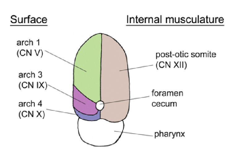
Primordia of developing tongue:
Lateral lingual swellings- Medial most parts of first pharyngeal (mandibular) arches proliferate to form two lateral swellings.
Tuberculum impar - Also formed by proliferation of median most part of 1st pharyngeal arches. It is median swelling which partially separates two lateral lingual swelling.

Hypobranchial eminence- another median swelling formed by mesoderm of 2nd, 3rd, and part of 4th pharyngeal arches. It divides into two parts:
Cranial part (copula) which is related to 2nd & 3rd arches.
Caudal part, which is related to 4th arch. It forms the epiglottis. Immediately behind this swelling is the laryngeal orifice, which is flanked by the arytenoid swellings.
Formation of tongue:
Anterior two-thirds of tongue [derived from first pharyngeal arch] -
➤ Two lateral lingual swelling increase in size, merge with each other, overgrow the tuberculum impar.
➤ The merged lateral lingual buds form the anterior two-thirds of the tongue.
➤ Fusion of the two lateral lingual buds is indicated by middle groove, the median sulcus of tongue.
Posterior one-thirds of tongue [formed by copula-cranial part of hypobranchial eminence] -
➤The 3rd arch mesoderm of copula grows over the 2nd arch mesoderm of copula and fuses with mesoderm of 1st arch.
➤ The V shaped groove separates the anterior two-thirds of the tongue from the posterior one third.

Most posterior part of tongue and epiglottis -
➤ Formed by caudal part of hypobrachial eminence derived from 4th arch mesoderm
Musculature of the tongue -
➤ Mainly derived from myoblasts originating in occipital somites. Tongue musculature is innervated by hypoglossal nerve.

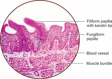
Histology of tongue:
- The bulk of tongue made up of striated muscles.
- The mucous membrane consists of a layer of connective tissue (corium), lined by stratified sqamous epithelium.
- On the oral part of the dorsum, it is thin, forms papillae & adherent to the muscles.
- On the pharyngeal part of the dorsum, it is rich in lymphoid follicles.
- On the inferior surface, it is thin and smooth. Numerous glands, both mucous & serous lie deep to the mucous membrane.
Taste buds: numerously present on side of vallate papillae, on wall of surrounding sulci, foliate papillae, and over the posterior one-third of tongue, sparsely on fungiform papillae, soft palate, epiglottis and pharynx.
No taste buds on mid-dorsal region of oral part of tongue.


Clinical anatomy:
- The undersurface of tongue & bulbar conjunctiva is good site for observation of jaundice.
- Carcinoma of the tongue is quite common. The affected side of tongue is removed surgically. all deep cervical lymph node also removed. [block dissection of neck because recurrence of malignant disease occurs in lymph nodes].
- Carcinoma of posterior one-third of tongue is more dangerous due to bilateral lymphatic spread.
- Lingual tonsil in posterior one-third of tongue forms part of waldeyer's ring.
- Referred pain felt in ear in diseases of posterior part of the tongue, as ninth nerve is common supply to both regions.
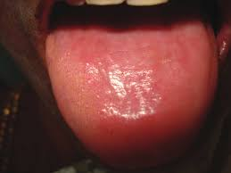
- In certain anemia tongue becomes smooth due to atrophy of filiform papillae.
- Sorbitrate taken sublingually for immediate relief from angina pectoris. It absorbs fast because of rich blood supply of the tongue & bypassing of hepatic circulation.
- Genioglossus is called 'safety muscle of the tongue', because if it is paralysed, tongue will fall back on oropharynx and block air passage.
- During anesthesia, tongue pulled forward to clear air passage.


Check below link for muscles of tongue
https://www.blogger.com/blog/post/edit/4923003805564511941/153432422799853043
Please let me know in the comment section if you have any doubts or you want me to upload any other topics.


No comments:
Post a Comment