Maxillary branch arises from middle of trigeminal ganglion and purely sensory in function.
Origin:
➤Leaves cranium through foramen rotundum. [Foramen rotundum located in greater wing of the sphenoid bone]
➤Crosses pterygopalatine fossa gives off branches to - sphenopalatine ganglion, posterior superior alveolar nerve, zygomatic branches.
➤Enters orbit through inferior orbital fissure, occupies infraorbital groove & becomes the infraorbital nerve, which course anteriorly into infraorbital canal.
➤Emerges on anterior surface of the face, through infraorbital foramen- divides into terminal branches, supplying skin of face, nose, lower eyelid, upper lip.

Branches:

Brief discussion of maxillary nerve branches:
Within cranium:
Middle meningeal nerve- provide sensory innervation to duramater.
Pterygopalatine fossa:
Zygomatic nerve:
It enters orbit through inferior orbital fissure and gives off branches:
➔Zygomaticotemporal branch- Sensory innervation to skin on side of forehead.
➔Zygomaticofacial- Skin on prominence of cheek.
Before leaving orbit: Zygomatic nerve sends a branch that communicates with lacrimal nerve of ophthalmic division.
It carries secretory fibre from sphenopalatine ganglion to lacrimal gland.
Pterygopalatine nerve:
Serve as a communication between pterygopalatine ganglion & maxillary nerve.
Branches:
1)Orbital branch:
supplies periosteum of orbit.
2)Nasal branch:
mucous membrane of superior & middle conchae, lining of posterior ethmoidal sinuses, posterior portion of nasal septum.
Course: passes across the roof of nasal cavity, where it lies between mucous membrane & periosteum of nasal septum.
➜Continues downward reaches floor of nasal cavity, and gives branches to anterior part of nasal septum and floor of nose.
➔Then enters incisive canal, through which it passes into oral cavity via incisive foramen located in midline of palate about 1cm posterior to maxillary central incisors.
➔Right & left nasopalatine nerve emerge together through incisive foramen- provide sensation to palatal mucosa in region of premaxilla.

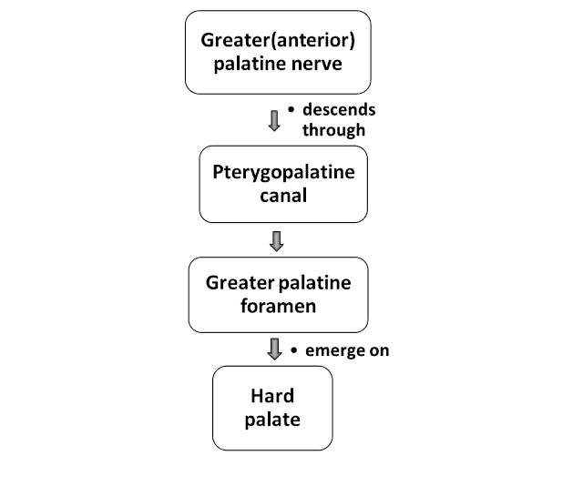
3)Palatine branches:
➔Greater (anterior) palatine nerve.
➔Lesser (middle & posterior) palatine nerve.
➣Middle & posterior palatine nerve emerge from lesser palatine foramen.
➣Middle palatine nerve innervates mucous membrane of soft palate.
➣Posterior palatine nerve innervates tonsillar region.
4)Pharyngeal branch:
Distributed to mucous membrane of nasal part of pharynx, posterior to auditory tube.
Posterior superior alveolar[PSA] nerve:
In pterygoplaltine fossa- PSA nerve descends from main trunk of maxillary division.
Two branch present on both side.
- One branch provide sensory innervation to buccal gingiva in maxillary molar region, adjacent facial mucosal surface.
- Other branch travel down posterior or posterolateral wall of maxillary sinus, provide sensory innervation to mucous membrane of sinus, alveoli, periodontal ligament, pulpal tissue of maxillary 1st, 2nd, 3rd molar.

Infraorbital canal:
Within infraorbital canal, maxillary division gives off two branches-
- Middle superior alveolar nerve.
- Anterior superior alveolar nerve.
Middle superior alveolar nerve:
Form part of superior dental plexus.

Provide sensory innervation to two maxillary premolars, mesiobuccal root of 1st molar, periodontal tissue, buccal soft tissue, & bone in premolar region.

Anterior superior alveolar nerve:
➤Large branch.
➤Gives out infraorbital nerve, descending within anterior wall of maxillary sinus, provide pulpal innervation to central, lateral incisor, canine, periodontal tissue, buccal bone & mucous membrane of these teeth.

Dental plexus:
Actual innervation of individual roots of all teeth, bone & periodontal structure in both maxilla & mandible derived from terminal branches of larger nerve in the region. These nerve network is called dental plexus.

Superior dental plexus:
Composed of small nerve fibre from 3 superior alveolar nerve.
3 types of nerve emerge from these plexus: Dental nerve, Interdental nerve & interradicular nerve.
- Dental nerve: enter a tooth through apical foramen, provide pulpal innervation of all tooth.
- Interdental nerve: (perforating branches) travel entire height of interradicular septum, providing sensory innervation to PDL of adjacent teeth through alveolar bone, innervate interdental papilla, buccal gingiva.
- Interradicular branch: travel entire height of interradicular or interalveolar septum, providing sensory innervation to PDL of adjacent root. Terminate in PDL at root furcation.
Branches on face:


Inferior palpebral branch:
Supplies skin of lower eyelid.
External nasal branch:
Supplies skin on lateral aspect of nose.
Superior labial branch:
Supplies skin & mucous membrane of upper lip.
Clinical anatomy:
In injury to Maxillary nerve- there is loss of sneeze reflex. This branch is the afferent path of sneeze reflex.
If you have any doubts related to this article, please let me know in the comment section.


No comments:
Post a Comment